TEMPORAL BONE ANATOMY
The temporal bone consists of five parts: the squamous, mastoid, petrous, tympanic and styloid portions. The squamous portion articulates with the occipital, parietal and sphenoid bones, and is part of the lateral wall of the middle cranial fossa and medial wall of the temporal fossa. The petrous is a quadrangular pyramid-shaped mass of the inner surface of the temporal bone, it extends to the petrous apex and contains the inner ear structures (vestibule, semicircular canals, cochlea) and virtually most neurovascular compartments (internal auditory canal, carotid canal and the jugular fossa); the apex of the pyramid rests on the clivus in the petro-occipital fissure. The tympanic bone forms the bulk of the ear canal and the middle ear space. It is separated anterior and internally from the petrous bone by the petrotympanic fissure, and anterior and externally from the squamous by the tympanosquamous suture. The mastoid portion contains the mastoid air cells, and articulates laterally with the parietal and occipital bones, and communicates with the nasopharynx through the Eustachian tube. There is continuity between the mastoid air cells and the petrous apex. The mastoid process forms a bony protrusion or eminence on the retro auricular region that protects the facial nerve, and it is not ossified at birth. The Körner's septum is part of the petrosquamous suture that runs posterolaterally through the mastoid air cells. It works as a barrier to the spread of infection from the lateral mastoid air cells to the medial ones. It also functions as a major surgical mark within the mastoid air cells. There is also a rudimentary temporal styloid, which originates in the second branchial arch and forms the posterior tympanum and the styloid process.
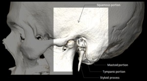
Fig. 1: Squamous, mastoid, petrous, tympanic and styloid portions of the temporal bone.
EAR
The outer and middle parts of the ear are responsible for transferring sound waves into the inner ear, which converts them into nerve impulses and records balance changes. External ear consists of the pinna, the external acoustic meatus and the outer wall of the tympanic membrane. Middle ear consists of three movable ossicles connecting the inner wall of the tympanic membrane and the oval window of the inner ear. Inner ear consists of the vestibulocochlear body in charge of both the hearing and the balance.
EXTERNAL EAR
The purposes of the external auditory meatus and canal are to collect and conduct the sound to the tympanic membrane as a resonant tube resulting in a gain from 10 to 20 dB.
The external auditory canal is 25 mm long and is filled with air in its normal state. It has two parts: the lateral third is made up of fibrocartilage with an incomplete ring of elastic cartilage measuring 8 mm long. It is lined by thick skin with hair follicles and glands. The two medial thirds are the bony portion, which is surrounded by the squamous, mastoid and tympanic segments of the temporal bone. It is lined with a thin layer of skin without follicles and glands. Anterior to the osseous part is the mandibular condyle, which however, is frequently separated from the cartilaginous part by a portion of the parotid gland. Posterior to the osseous part are the mastoid air cells, separated from the meatus by a thin layer of bone.
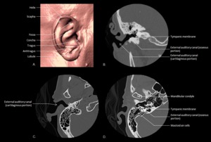
Fig. 2: External ear anatomy: The pinna (A), the external acoustic meatus and the outer wall of the tympanic membrane in coronal (B) and axial (C, D) views.
MIDDLE EAR
It is also filled with air, which is transmitted from the nasal part of the pharynx through the Eustachian tube. It contains a string of moving bones, the ossicles, whose primary role is to mechanically transmit and amplify vibrations received by the tympanic membrane to the cochlea. The tympanic cavity is divided into 3 regions; epitympanum, mesotympanum and hypotympanum. The epitympanum contains the most of the structures.
The lateral wall is formed mainly by the tympanic membrane that separates the tympanic cavity from the bottom of the external acoustic meatus. It is a thin, semitransparent membrane, nearly oval in form, somewhat broader above than below, and directed very obliquely downward and inward. The greater part of its circumference is thickened, and forms a fibrocartilaginous ring which is fixed in the scutum and annulus at the inner end of the meatus. The manubrium of the malleus is firmly attached to the medial surface of the membrane as far as its center, which it draws toward the tympanic cavity; the lateral surface of the membrane is thus concave, and the most depressed part of this concavity is called the umbo.
The tegmental roof has two parts; the tegmen tympani, which is made by a thin plate of bone and separates the cranial and tympanic cavities; and the arcuate eminence which is the bony prominence on the superior semicircular canal, is a surgical mark when the neurosurgeon cuts along the floor of the middle cranial fossa.
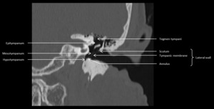
Fig. 3: Middle ear anatomy: Epitympanum, mesotympanum, hypotympanum, tegmen tympani, scutum, lateral wall in coronal view.
The jugular wall or floor is narrow, and consists of a thin plate of bone (fundus tympani) which separates the tympanic cavity from the jugular fossa.
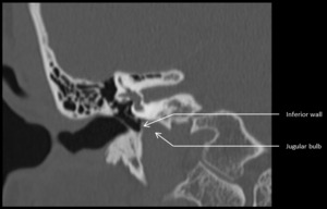
Fig. 4: Inferior wall and jugular bulb in coronal view.
There are several marks at the medial wall. The oval window is a reniform opening leading from the tympanic cavity into the vestibule of the internal ear. In the recent state it is occupied by the base of the stapes, the circumference of which is fixed by the annular ligament to the margin of the foramen. The round window is situated below and a little behind the oval window, from which it is separated by a rounded elevation, the promontory and leads into the cochlea of the internal ear.
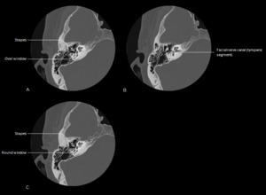
Fig. 5: Middle ear anatomy: Stapes, facial nerve canal, oval and round window in axial view (A, B, C).
The promontory (promontorium) is a rounded hollow prominence, formed by the projection outward of the first turn of the cochlea; it is placed between the windows, and is furrowed on its surface by small grooves, for the lodgement of branches of the tympanic plexus.
The prominence of the facial canal indicates the position of the bony canal in which the facial nerve is contained; this canal traverses the labyrinthic wall of the tympanic cavity above the oval window.
The aditus ad antrum is located at the upper posterior wall and connects the epitympanum of the middle ear cavity with the mastoid antrum. The bottom portion of the posterior wall consists of three major structures: The facial recess, pyramidal eminence and sinus tympani.
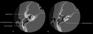
Fig. 6: Middle ear anatomy: Facial recess, pyramidal eminence, sinus tympani and aditus ad antrum in axial views (A, B).
The anterior wall is wider above than below; it corresponds with the carotid canal, from which it is separated by a thin plate of bone perforated by the tympanic branch of the internal carotid artery, and by the deep petrosal nerve.
At the upper part of the anterior wall are the orifice for the Tensor tympani muscle and the tympanic orifice of the auditory tube, separated from each other by a thin horizontal plate of bone. These canals run from the tympanic cavity forward and downward to the retiring angle between the squama and the petrous portion of the temporal bone.
The orifice for the Tensor tympani extends on to the labyrinthic wall of the tympanic cavity and ends immediately above the oval window. The auditory tube (tuba auditiva; Eustachian tube) is the channel through which the tympanic cavity communicates with the nasal part of the pharynx. It is formed partly of bone, partly of cartilage and fibrous tissue.
OSSICLES
The normal ossicular chain consists of the malleus, incus and stapes. The first is attached to the tympanic membrane, and it consists of a head, neck, and three processes; the manubrium, the anterior and the lateral (short) processes. The head is the large upper extremity of the bone; and articulates posteriorly with the incus, being free in the rest. The manubrium is connected by its lateral margin with the tympanic membrane.
The incus consists of a body and the short, long and lenticular processes. The body has a deeply concavo-convex facet on its anterior surface, which articulates with the head of the malleus. The long process ends in a rounded projection, the lenticular process, which is tipped with cartilage, and articulates with the head of the stapes.
The stapes consists of a head, neck, anterior crus and posterior crus, and the footplate. The head (Capitulum) presents a depression, which is covered by cartilage, and articulates with the lenticular process of the incus. The neck, the constricted part of the bone succeeding the head, gives insertion to the tendon of the stapedius muscle. The two crura diverge from the neck and are connected at their ends by the footplate, which is fixed to the margin of the oval window by the annular ligament.
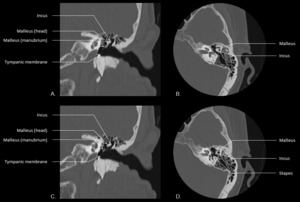
Fig. 7: Middle ear anatomy: Ossicles and tympanic membrane in coronal (A, C) and axial (B, D) views.
INNER EAR
The inner ear consists of a membranous (endolymphatic) labyrinth containing the functional sensory epithelium surrounded by an osseous labyrinth with an interposed perylimphatic labyrinth.
The membranous labyrinth consists of the utricle, saccule, semicircular ducts, cochlear duct, and endolymphatic duct and sac. All of these structures contain endolymph. The osseous labyrinth is the bony shell that surrounds the membranous labyrinth. The utricle and saccule (membranous labyrinth) are house within the vestibule (bony labyrinth), the semicircular duct (membranous labyrinth) within the semicircular canals (bony labyrinth), and the endolymphatic duct (membranous labyrinth) within the proximal vestibular aqueduct (bony labyrinth). The perilymphatic labyrinth is interposed between the membranous and bony labyrinth.
The vestibule is the central, rounded portion of the osseous labyrinth. In its lateral or tympanic wall is the oval window, closed, in the fresh state, by the base of the stapes and annular ligament. It is continuous anteroinferiorly with the cochlea and posteriorly with the semicircular canals and the vestibular aqueduct. Within the osseous vestibule the membranous labyrinth does not quite preserve the form of the bony cavity, but consists of two membranous sacs, the utricle, and the saccule.
The vestibular aqueduct is a tubular structure that arises from the vestibule and runs along the posteroinferior aspect of the petrous temporal bone. It contains the endolymphatic duct and sac, which are connected to the utricle and saccule of the vestibule. The vestibular aqueduct is diagonally oriented in relation to the direction of the IAC. The aqueduct, which normally measures less than 1.5 mm in diameter, approximates the size of the posterior semicircular canal, which runs anterior and parallel to the aqueduct.
The cochlea forms the anterior part of the labyrinth, is conical in form, and placed almost horizontally in front of the vestibule. Its base corresponds with the bottom of the internal acoustic meatus, and is perforated by numerous apertures for the passage of the cochlear division of the acoustic nerve. It consists of 2½ to 2 ¾ turns, and it involves a conical shaped central axis, the modiolus; and of a delicate lamina, the osseous spiral lamina that projects outward from the modiolus.
The cochlear aqueduct is a narrow canal which runs towards the cochlea in almost the same direction as the inner auditory canal, but situated more caudally. It extends from the basal turn of the cochlea to the subarachnoid space adjacent to the jugular foramen.
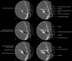
Fig. 8: Inner ear anatomy: Superior, lateral and posterior semicircular canals; internal auditory canal, labyrinthine portion CN7 canal, vestibule, bony vestibular aqueduct, cochlea, facial nerve and tensor tympani muscle in axial views (A - F).
0 Response to "Ct Imaging of Temporal Bone Making Easy What Used Ot Be Hard"
Post a Comment